Veterinary Radiology and Ultrasonography Mentorship
Module 4: Advanced Ultrasound, Guided Procedures, Ocular and Neck Ultrasound


Every day in practice, you make decisions from images.
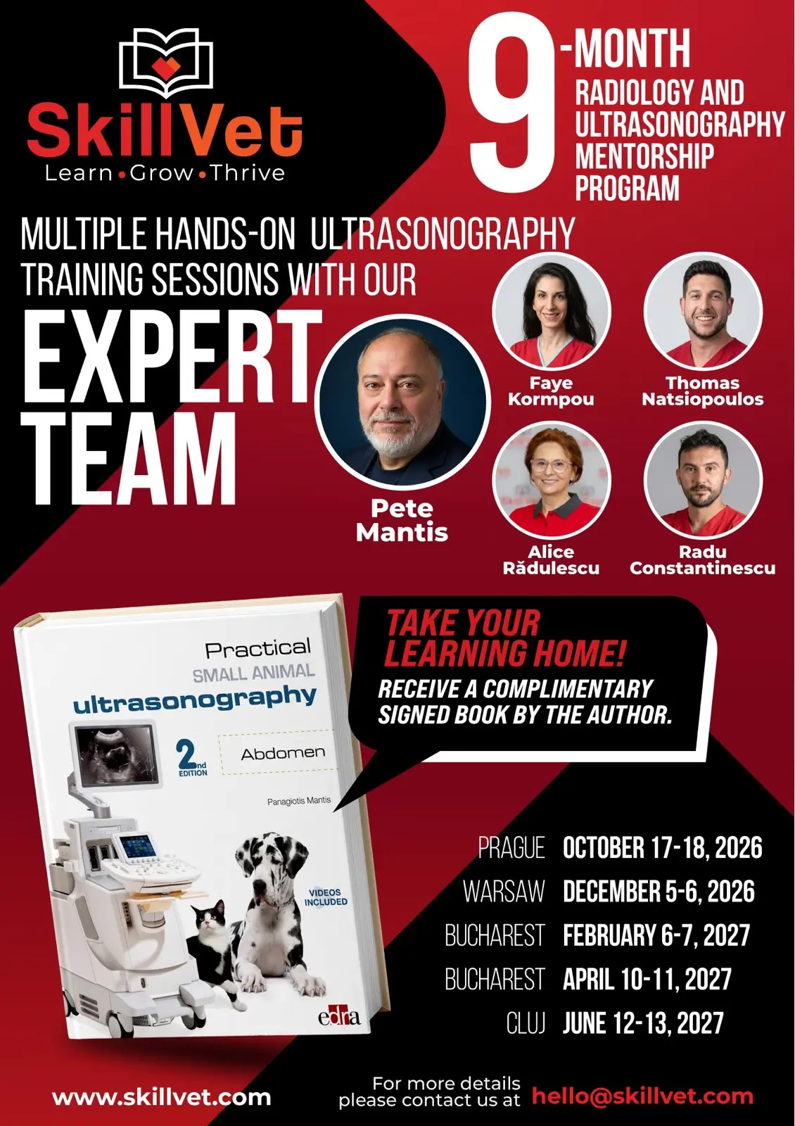
The Veterinary Radiology and Ultrasonography Mentorship is a 9-month program — five modules across four European cities, a personal Diplomate mentor throughout, and a final assessed certification.
You can Register here to this event together with all it's modules listed below, part of this mentorship program.
19+ hours
of hands-on practice
- October 17-18, 2026 - Prague
- December 5-6, 2026 - Warsaw
- February 6-7, 2027 - Bucharest
- April 10-11, 2027 - Bucharest
- June 12-13, 2027 - Cluj
Complete Abdominal Scanning, FNA/Biopsy Technique, and Beyond the Abdomen
Abdominal scanning established, this module extends into the skills many practitioners have never been formally taught: ultrasound-guided FNA and biopsy, ocular ultrasound, and neck and thyroid scanning. Hands-on throughout.
Course description
Diagnostic imaging is part of daily small-animal practice. Whether you interpret radiographs regularly, perform abdominal ultrasound, or want to build those skills from the ground up — the question is the same: how do you move from doing it to doing it well, consistently, in any case that walks through your door? This program is designed to answer that question.
The program is intentionally structured as a longitudinal mentorship rather than a short isolated workshop. Its aim is to move participants from a basic or inconsistent imaging approach toward a more systematic, reproducible, and clinically relevant method for evaluating imaging cases in daily practice. It runs from October 2026 through June 2027 across five European teaching weekends in Prague, Warsaw, Bucharest, and Cluj-Napoca.
What makes this program distinctive is the integration of radiology and ultrasonography into a single, coherent diagnostic imaging pathway. In clinical practice, imaging modalities are rarely interpreted in isolation — thoracic radiographs, abdominal radiographs, musculoskeletal studies, abdominal ultrasound, neck and ocular ultrasound, and ultrasound-guided sampling are frequently combined to answer the same clinical question. The program teaches participants not only how to recognize imaging abnormalities, but how to place those findings within a rational diagnostic and clinical decision-making framework.
The radiology component begins with structured thoracic and abdominal interpretation, progresses through musculoskeletal radiology, and is reinforced through repeated case sessions. The ultrasound component covers physics, artifacts, machine optimization, and systematic abdominal scanning — then extends to guided procedures, ocular, neck, thyroid, and tendon ultrasound. Each module builds on the one before it, and between-module mentorship ensures that learning is applied to real clinical work — not held in reserve until the next weekend.
The program is designed for veterinarians who want to build real diagnostic confidence, not only theoretical familiarity. Structured reporting, clinical reasoning, recognition of examination limitations, and knowing when to refer are embedded throughout the curriculum.
Teaching philosophy
Diagnostic imaging is a skill set that requires pattern recognition, technical acquisition, anatomical knowledge, clinical reasoning, and disciplined reporting. Short courses can provide useful exposure, but they rarely provide the repetition, supervision, and feedback needed to change day-to-day clinical practice. This mentorship was designed to do exactly that.
The program uses a spiral learning model. Core principles are introduced early, revisited repeatedly, and applied with increasing complexity. Thoracic radiology establishes an interpretive discipline in Module 1. That same discipline is applied to musculoskeletal cases in Module 2 and to ultrasonography from Module 3 onward. By the final module, participants are expected to integrate radiology and ultrasound findings in case discussions and examinations — using a single, consistent clinical reasoning framework across both modalities.
A central pillar of the program is deliberate practice. Ultrasonography is highly operator-dependent. Competence develops through repeated supervised scanning opportunities — acquiring the same structures, optimizing the same image planes, receiving immediate correction, and refining technique across multiple sessions with different patients. The course provides a high volume of hands-on ultrasound training in small groups with a high instructor-to-participant ratio, because the aim is not to demonstrate a technique once, but to allow participants to perform it, make mistakes safely, correct those mistakes, and build a more reliable scanning pattern.
Radiology teaching is similarly designed around active interpretation. Participants are not shown examples of disease — they are asked to apply a systematic method, describe findings in appropriate terminology, identify the primary lesion pattern, build prioritized differentials, and consider what additional imaging or clinical information is required. This trains the kind of reasoning that produces useful clinical reports, not isolated pattern recognition.
The program's core commitment is clinical relevance. Participants leave each module with skills that can be applied in practice the following week — not only in the structured environment of a course.
Learning objectives
By the end of Module 4, participants will be able to:
- Improve confidence in identifying and scanning the left and right adrenal glands.
- Perform a more complete and consistent abdominal ultrasound examination.
- Understand the indications, contraindications, safety requirements, and documentation needs for ultrasound-guided fine needle aspirates and biopsies.
- Recognize basic ocular ultrasound anatomy and understand common indications for ocular ultrasound.
- Identify thyroid and parathyroid region anatomy and understand how ultrasound contributes to clinical decision-making in neck disease.
- Perform a basic ultrasound assessment of the neck, including salivary glands, laryngeal/tracheal region, and regional lymph nodes.
- Integrate ultrasound findings into clinical recommendations while recognizing when additional imaging or specialist review is appropriate.
Educational objectives
By the end of the full mentorship, participants will be able to:
- Interpret thoracic radiographs using a structured, clinically relevant approach including image quality assessment, anatomical review, lesion localization, pattern recognition, and clinical prioritization
- Recognize and differentiate key thoracic abnormalities: lung patterns, pleural disease, mediastinal abnormalities, chest wall and diaphragmatic lesions, thoracic masses, and cardiac radiographic changes
- Perform systematic interpretation of abdominal radiographs, including evaluation of serosal detail, organ size, organ position, gastrointestinal gas patterns, urinary and reproductive tract changes, and abdominal masses
- Identify common musculoskeletal radiographic abnormalities including developmental disease, mature-patient disease, trauma, and aggressive versus non-aggressive bone lesions
- Apply a logical, imaging-based diagnostic approach to complex cases, integrating signalment, clinical history, examination findings, radiology, and ultrasonography
- Understand ultrasound physics, artifacts, machine controls, probe selection, patient positioning, and image optimization principles
- Perform a systematic abdominal ultrasound examination in dogs and cats, including the liver, gallbladder, spleen, gastrointestinal tract, pancreas, kidneys, urinary bladder, reproductive tract, adrenal glands, lymph nodes, peritoneum, and retroperitoneum
- Perform selected non-abdominal ultrasound examinations including ocular, thyroid and parathyroid, neck (larynx, trachea, salivary glands), regional lymph node, and tendon ultrasound
- Understand indications, limitations, safety considerations, and practical technique for ultrasound-guided fine needle aspirates and biopsies
- Integrate radiographic and ultrasonographic findings into structured diagnostic imaging reports that are clinically useful for general practice and referral communication
- Recognize when image quality, case complexity, or patient safety requires referral, repeat imaging, advanced imaging, or specialist review
- Apply ethical principles, patient safety considerations, and evidence-based reasoning when performing diagnostic imaging and ultrasound-guided procedures
Between-module mentorship
Learning in diagnostic imaging does not stop when the weekend ends. Each participant in this program is mentored continuously between modules — because practical improvement occurs when skills are applied repeatedly to real clinical material in the weeks that follow a module, not only during it.
Between modules, participants are encouraged to submit radiographic cases, ultrasound images, short video clips, draft reports, and interpretation exercises. Submitted material may include thoracic, abdominal, or musculoskeletal radiographs, abdominal ultrasound stills or cine loops, or structured written reports. Faculty feedback focuses on image quality, completeness of examination, lesion description, prioritization of findings, differential diagnosis, and clinical recommendations.
The mentorship is designed to help participants recognize strengths and weaknesses in their own approach, improve future image acquisition, and develop a more disciplined method of interpretation. When clinically significant uncertainty is present, participants are guided toward referral or specialist review.
Participants may also take part in structured group discussions or one-to-one mentoring conversations to clarify concepts, work through difficult cases, prepare for the next module, and receive guidance on personal learning goals. This structure supports continuity, accountability, and the kind of progressive improvement that is difficult to achieve through a series of disconnected workshops.
Evaluation and certification
Assessment is designed to evaluate both theoretical understanding and practical competence. Diagnostic imaging cannot be assessed through knowledge testing alone — participants must also demonstrate the ability to acquire images, recognize anatomy, describe abnormalities, interpret cases, and communicate findings in a clinically useful way.
Formative assessment occurs throughout the program through case discussions, practical scanning feedback, between-module homework, report review, quizzes, and instructor observation during scanning sessions. This ongoing feedback allows faculty to identify recurring difficulties early and provide targeted correction.
The final assessment is summative and takes place during Module 5 in Cluj-Napoca. It consists of a final theoretical examination (60% of the overall assessment) and a final practical examination (40%), during which participants rotate through assessment stations.
Participants who successfully complete all requirements and pass the final assessments are awarded the SkillVet Certificate in Veterinary Radiology and Ultrasonography. The certificate reflects successful completion of a structured postgraduate continuing education mentorship; it does not imply specialist status or board certification.
Instructional methods
The program combines multiple instructional methods, because diagnostic imaging competence requires knowledge, interpretation, practical acquisition, and clinical judgment — and no single format develops all four.
Theoretical lectures provide structured frameworks rather than isolated facts. In radiology, participants learn how to approach images in a repeatable order and identify the primary lesion pattern. In ultrasonography, lectures introduce anatomy, expected normal appearance, artifacts, and common abnormalities before participants scan — so they know what they are looking for and how artifacts may mislead them.
Case discussions train clinical reasoning. Participants are asked to describe findings before assigning a diagnosis, to separate observation from interpretation, and to build a prioritized differential list. This develops the kind of report that is useful to clinicians and clients rather than vague or overly speculative.
Practical ultrasound sessions are designed around ergonomics, probe handling, image optimization, patient positioning, and systematic organ coverage. Instructors provide immediate correction — because small changes in probe angle, pressure, rotation, depth, or gain alter the image significantly, and participants need to understand why, not only what to do differently.
Between-module mentorship activities include case submissions, interpretation exercises, quizzes, reflective assignments, and optional one-to-one support for participants with specific learning goals.
SkillVet is independent and vendor-neutral. This program does not depend on any specific ultrasound machine, radiographic system, software platform, or commercial product. When equipment features are discussed, they are explained through general imaging principles that apply across different clinical settings. No manufacturer sponsorship determines the curriculum.
Expected outcomes
Upon completing the Veterinary Radiology and Ultrasonography Mentorship Program, graduates will be able to:
- Use a structured method to interpret thoracic, abdominal, and musculoskeletal radiographs
- Recognize common thoracic, abdominal, and musculoskeletal imaging patterns and describe them accurately
- Perform a more complete and systematic abdominal ultrasound examination in dogs and cats
- Recognize normal ultrasound anatomy and common abnormalities of the major abdominal organs
- Perform selected ocular, neck, thyroid/parathyroid, lymph node, and tendon ultrasound examinations at an introductory-to-intermediate level appropriate to this program's scope
- Understand how to perform ultrasound-guided sampling safely and recognize when such procedures are or are not appropriate
- Produce clear, structured diagnostic imaging reports that distinguish findings, interpretation, differentials, limitations, and recommendations
- Integrate radiology and ultrasonography findings into rational clinical decision-making
- Recognize when image quality, case complexity, or patient safety requires repeat imaging, referral, advanced imaging, or specialist interpretation
- Apply imaging knowledge in daily small-animal practice with greater confidence, consistency, and professional responsibility
Faculty

Dr. Pete Mantis, DVM, DipECVDI, FHEA, PhD, FRCVS
Program Head and Lead Mentor
European and RCVS Specialist in Veterinary Diagnostic Imaging and Head of Diagnostic Imaging at Dick White Referrals. Honorary Associate Professor at the University of Nottingham and author of Practical Small Animal Ultrasonography: Abdomen. Dr. Mantis serves as Program Head and Lead Mentor, providing academic oversight, curriculum leadership, and quality assurance throughout the mentorship.

Dr. Faye Kormpou, DipECVDI
Instructor
Diplomate of the European College of Veterinary Diagnostic Imaging and Staff Clinician in Small Animal Diagnostic Imaging at the Royal Veterinary College. Dr. Kormpou also provides teleradiology services to practices across the UK and internationally, bringing a sharp perspective on the full range of imaging presentations encountered in daily general practice.
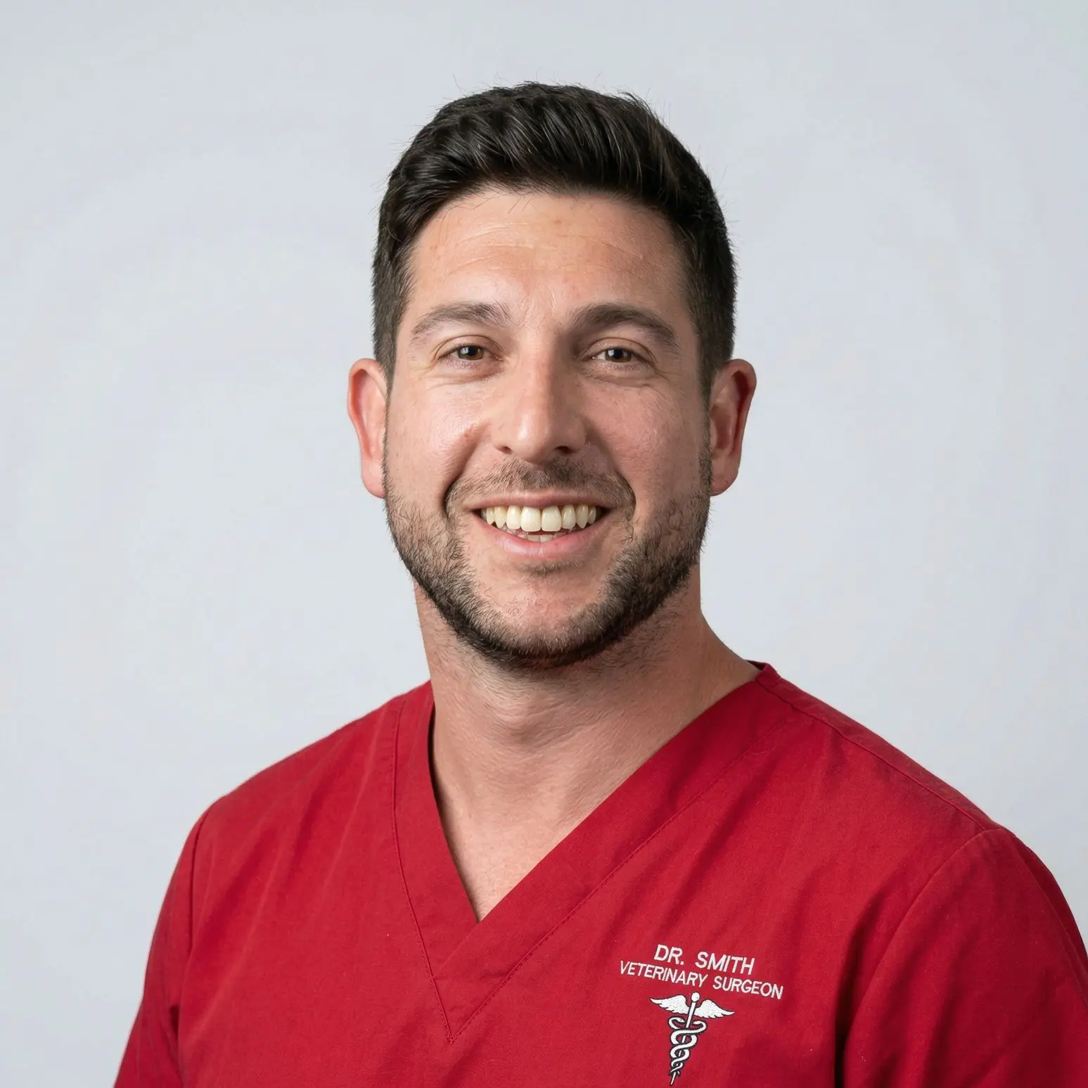
Dr. Thomas Natsiopoulos, DVM, MRCVS, DipECVDI
Instructor
EBVS European Specialist in Veterinary Diagnostic Imaging at Lumbry Park Veterinary Specialists. Dr. Natsiopoulos has particular interests in musculoskeletal imaging, neuroimaging, and interventional procedures. He has been part of the SkillVet community since its early days and is invested in the kind of education that produces independently capable veterinarians.

Dr. Alice Rădulescu, DVM, PVP
Instructor - Ultrasound Practical Teaching, Scanning Supervision, and Clinical Mentorship
Founder of A&A Medical Vet in Bucharest and President of the Romanian Association of Veterinary Cardiologists, with over 25 years of clinical experience in small animal cardiology and ultrasound. Dr. Rădulescu leads the practical ultrasound teaching for the Bucharest modules and is the reason this program has a home in Romania.
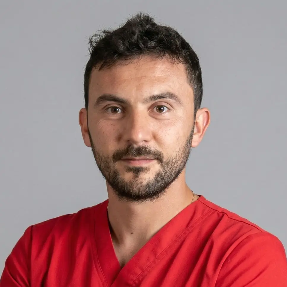
Dr. Radu Constantinescu, DVM, PhD
Instructor, ECVDI Resident
Romanian veterinarian and ECVDI Resident at Dick White Referrals, with a PhD in Veterinary Clinical Sciences focused on renal ultrasonography. He has published on ultrasonography, radiology, CT, MRI, and congenital vascular abnormalities, and brings over a decade of clinical imaging experience - from private practice in Bucharest to referral-level teaching in the UK.
Is this program right for you?
This program is designed for qualified veterinarians working in small-animal general or referral practice who want to build a more structured, reliable approach to diagnostic imaging.
It is suitable for you if:
- You perform or interpret radiographs regularly and want to develop a more systematic, reproducible method
- You are beginning to learn small-animal ultrasound and want a supervised, progressive pathway — not a single introductory workshop
- You have some ultrasound experience but want to expand your range, improve image quality, and gain confidence in more challenging regions
- You want a certification that reflects real, assessed competence — not only attendance
- You want continuity of learning with a personal mentor throughout, not isolated modules with no connection between them
This program is not intended as a substitute for a diagnostic imaging residency or specialist training. It is continuing education at a level appropriate for general and advanced small-animal practice — structured to improve confidence, consistency, and clinical responsibility in your day-to-day imaging work.
Prerequisites: Degree in veterinary medicine; active clinical practice involving small animals. No prior formal imaging training is required, but participants should be comfortable working with live patients in a small-group clinical teaching environment.
Conclusion
If you regularly pick up a radiograph or reach for an ultrasound probe in your practice, this program is built for you. Not for the abstract theory. For what happens after you complete the exam — when you need to make a decision, write a report, and know whether to refer.
Five weekends across nine months. A personal mentor throughout. Small groups, direct feedback, and the kind of progressive skill development that doesn't disappear between modules.
Registration fees
- Registration fee - 4,600€ (21% VAT included)
The price includes VAT
Romanian
VAT (21%) is legally required for all participants, regardless of country of
origin or VAT registration status. This is a legal obligation under EU and
Romanian tax law for in-person educational events held in Romania; it cannot be
waived or exempted.
What's included: Nine months of structured mentorship
- Five in-person modules across four cities
- A personal Diplomate mentor between modules
- Access to theory lecture recordings
- The final theoretical and practical examination
- Your SkillVet Certificate in Veterinary Radiology and Ultrasonography
- A personal copy of Practical Small Animal Ultrasonography: Abdomen by Dr. Pete Mantis
- Lunch, coffee, and water on both days of each module
Not included: Accommodation, travel, and evening meals
Limited places — registrations are on a first-come, first-served basis.
Apply Now
FAQ
Registration covers the full 9-month program, all 5 modules. Individual modules are not available separately.
The registration fee covers all course materials, access to theory lecture recordings, lunch, coffee, and water on both days of each module, and a personal copy of Practical Small Animal Ultrasonography: Abdomen by Dr. Pete Mantis. Accommodation, travel, and evening meals are not included.
English
Participants who miss a module for reasons of force majeure receive access to the theory lecture recordings for that module. Practical skills from a missed module can be recovered during a subsequent module by arrangement. Attendance at all five modules is expected; please contact us before registering if you have a known scheduling conflict.
Yes. Installment plans are available for a maximum of four payments. To request a plan, email hello@skillvet.com with the number of installments you need; the team will set up a payment schedule for you.
Yes. Contact us at hello@skillvet.com.
Cancellations submitted more than 30 days before the program start date are eligible for a full refund. Cancellations received within 30 days of the program start date are not eligible for a refund. To cancel, contact hello@skillvet.com.
Accommodation, flights, and evening meals are not included in the registration fee and are the responsibility of each participant.
No scholarships are currently available.
You have to register through main (first) event part of this mentorship program.