Milo was a young rescue dog recently imported from Eastern Europe. His owners noticed that he was quieter than expected, tired easily on walks, and had developed a dry, harsh cough over the previous 24 hours. On clinical examination, he was tachypnoeic and mildly lethargic. A loud grade V/VI left basilar systolic murmur was auscultated, and his rhythm was irregular. Because of the travel history and respiratory signs, heartworm disease was initially considered, but thoracic radiographs were performed to assess cardiac size, pulmonary vasculature, and possible pulmonary disease.
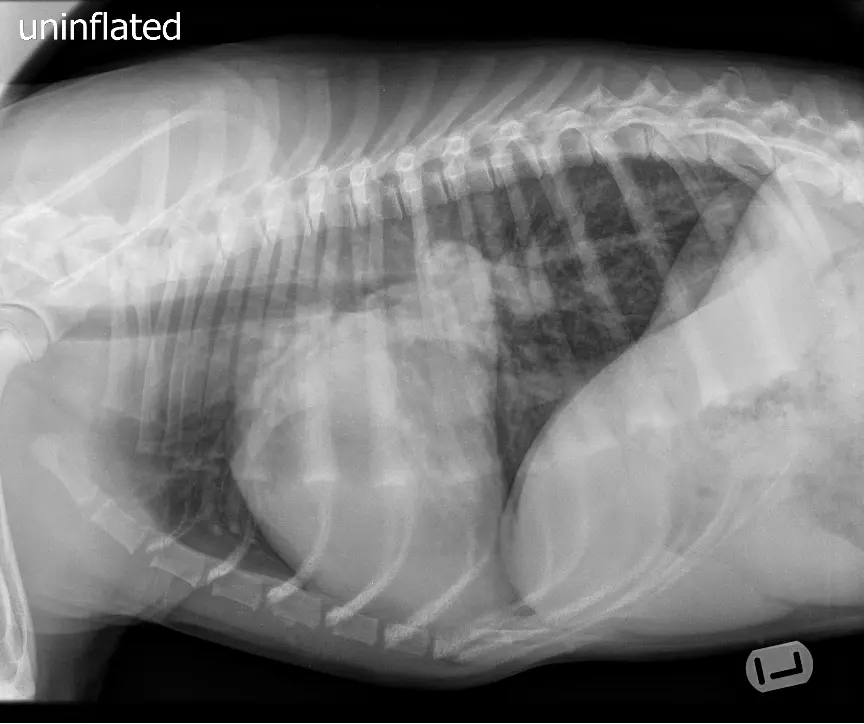
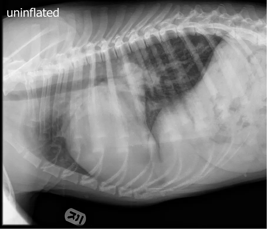
Thoracic radiographs showed marked cardiomegaly, with a VHS just under 12. The left atrium was severely enlarged, displacing the mainstem bronchi and extending dorsally to the carina. Both the pulmonary arteries and veins were enlarged, with the veins slightly larger than the arteries. There was also a mild generalised bronchointerstitial lung pattern, slightly more pronounced than expected for a patient of this age.
The radiographic interpretation was most consistent with left-sided cardiomegaly and pulmonary overcirculation. Because both the pulmonary arteries and veins were enlarged, this was considered more compatible with true pulmonary overcirculation rather than simple passive congestion from left-sided heart failure. The main congenital differentials included a patent ductus arteriosus, a large ventricular septal defect, or an atrial septal defect. A mild component of pulmonary oedema was also considered possible.
Milo was referred for echocardiography, which confirmed a haemodynamically significant left-to-right shunting congenital defect, most consistent with a patent ductus arteriosus. Given the severity of the left-sided volume overload, closure was recommended. He underwent minimally invasive occlusion using an appropriate vascular occlusion device. Peri-procedural monitoring was uneventful, and repeat echocardiography demonstrated successful closure with only trivial residual flow.
Over the following weeks, Milo’s respiratory rate normalised, his cough resolved, and his exercise tolerance improved dramatically. His owners reported that he became more playful and energetic at home. Follow-up imaging showed improvement in pulmonary vascular congestion and progressive reduction in left atrial and left ventricular size, consistent with reverse remodelling after successful closure of the shunt.
This case highlights how thoracic radiographs can raise strong suspicion for congenital overcirculation before echocardiography. In a young patient with a loud basilar murmur, marked left-sided cardiomegaly, and enlargement of both pulmonary arteries and veins, a significant left-to-right shunt should be high on the differential list.